Treatment of Congenital Urinary Tract Abnormalities in Children
Birth defects are a major concern for parents, and they believe that there is no cure. However, with modern technologies, treatment is possible.

Treatment of Congenital Urinary Tract Abnormalities in Children
Congenital urinary tract abnormalities in children are a significant concern for parents, as they can affect a child's ability to urinate and eliminate toxins from the body. They can also cause serious complications, such as kidney damage and impaired growth. Therefore, early diagnosis and intervention are crucial. Now, with the expertise of Dr. Enmar Mohamed Habib, Professor of Urology and Pediatric Urology at Cairo University and Fellow of McGill University, Canada, congenital malformations in children can be treated with the latest methods and techniques.
What are congenital urinary tract abnormalities in children?
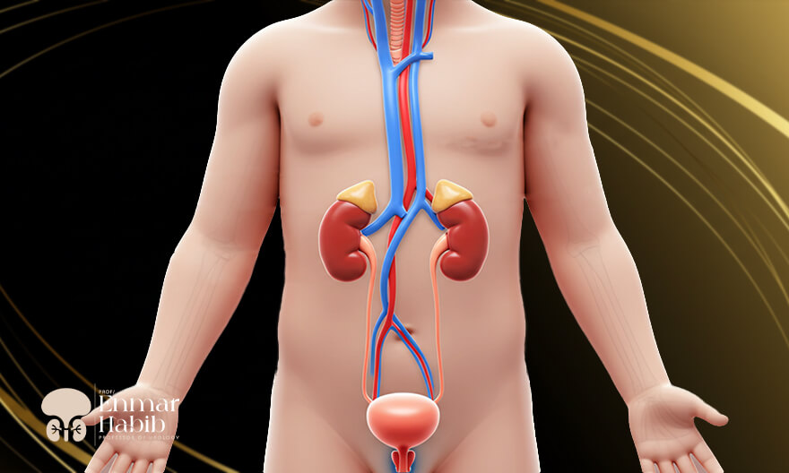
• Congenital urinary tract abnormalities are birth defects that affect the shape and function of the kidneys, urinary tract, and genitourinary system. Congenital abnormalities are deformities that occur during fetal development or are discovered early in a child's life.• Normal infants are born with kidneys that filter waste products and excess fluids from the blood. The kidneys also produce hormones that help strengthen bones, produce red blood cells, and control blood pressure.
• The body's waste products are eliminated in the form of urine from the kidneys to the bladder through the ureters (a muscular tube that carries urine from the kidneys to the bladder). Then the urine exits the bladder through the urethra (the urinary passage) to the outside of the body. Therefore, any abnormality in the function and shape of the kidneys or failure to pass urine normally is considered a congenital defect.
• Most congenital abnormalities are usually detected during pregnancy by an obstetrician through ultrasound imaging, while others are detected after birth.
What are the causes of congenital urinary tract abnormalities in children?
All parents wonder what causes congenital urinary tract abnormalities in their children. There is no known specific cause of malformations in general. However, many scientists believe that the occurrence of malformations is a combination of environmental factors and genetic mutations:• Consanguinity (marriage between relatives): Consanguineous marriage is one of the most likely causes of congenital malformations of the urinary system.
• Genetic mutations: Some genetic mutations, such as defects in certain genes that play a significant role in kidney and urinary tract formation, lead to congenital malformations.
• Vitamin and mineral deficiencies: Lack of crucial elements during pregnancy, such as folate and iron, is a major factor in urinary tract abnormalities.
Symptoms of congenital urinary tract abnormalities in children:
Most urinary tract malformations are detected during pregnancy. Therefore, when a malformation is identified, doctors closely monitor the pregnancy for signs of insufficient amniotic fluid, as this fluid is primarily made up of the urine produced continuously by the fetus after the 20th week of pregnancy.If congenital urinary tract malformations are not detected before birth, the early stages of a child's life may include the following symptoms:
• Recurrent urinary tract infections (sometimes presenting as an unexplained fever).
• Abdominal swelling (due to incomplete bladder emptying).
• Nausea and vomiting.
• Urinating more or less than usual.
• Blood in the urine (hematuria).
• Failure of the child to grow as expected.
Parents should carefully observe their children in the postnatal period to note any symptoms that may indicate urinary tract issues. Recognizing the symptoms that indicate an issue with the genitourinary system in children is quite challenging. Therefore, it is important to consult the best pediatric urologist, Dr. Enmar Mohamed Habib, to accurately diagnose the problem and treat congenital abnormalities safely and effectively.
Types of congenital urinary tract abnormalities in children and their treatment:
Abnormalities can occur anywhere in the urinary system. Some of these include:1. Urethral abnormalities (Epispadias and Hypospadias):
Congenital malformations of the urethra (the tube that carries urine from the bladder to the outside of the body) in males usually involve abnormalities in the anatomy or structure of the penis. However, in girls, urethral malformations can occur without abnormalities in the female reproductive system. Urethral abnormalities vary and include the following:
Epispadias:
• This is a rare congenital defect that a child is born with. Epispadias is more commonly seen in males, while it is rare in females. It occurs when the urethra does not fully develop into a complete tube, causing urine to exit the body from an abnormal location.• In males with epispadias, the urethral opening may be located on the top of the penis, along the shaft, or near the pubic area rather than at the tip of the penis.
• In females, epispadias results in the urethral opening in the abdomen rather than between the clitoris and the labia.
• Epispadias is often associated with a condition called bladder exstrophy, a rare birth defect in which the bladder is located outside the abdomen instead of inside.
• Epispadias are classified based on the precise location of the urethral opening, as the position directly impacts the ability of the bladder to store urine. The closer the opening is to the base of the penis, the more likely it is to affect the bladder sphincter, impairing the ability to control urination.
Treatment of Epispadias:
The treatment is by surgical intervention using the most advanced methods and techniques with the skills of Dr. Enmar Mohamed Habib. Surgery is recommended after the age of 6 months, as it helps achieve better results, especially regarding urinary control. The surgery is based on repositioning the urethral opening in the right place and correcting the deformity of the reproductive system's appearance.
Hypospadias:
• Hypospadias occurs when the urethra fails to form, and the urethral canal does not fuse properly, most commonly in males. In cases of hypospadias, the foreskin (the skin covering the head of the penis) does not develop correctly, so it does not form a complete circle but instead appears as a covering on the top part of the penis, leaving the underside of the penis uncovered.• In this condition, the urethral opening is located along the underside of the penis, at the junction between the penis and the scrotum (the skin pouch that holds the testes), between the folds of the scrotum, or in the perineal area (near the anus), rather than at the tip of the penis, where both urine and semen are normally expelled.
• Hypospadias may be associated with abnormalities in the appearance of the penis, such as a downward-curved penis, as well as skin irritation in the surrounding area. Without treatment, hypospadias can lead to urinary difficulties and infertility.
Treatment of Hypospadias:
• Surgical intervention is the best treatment for hypospadias. The surgery is typically performed between the ages of 6 and 12 months. Dr. Enmar Habib repositions the urethra to its natural location. If the urethra is underdeveloped, skin grafts may be used to reconstruct the urethral canal. The procedure may also involve correcting any curvature of the penis to make it straight.• In some cases, a temporary catheter may be placed in the urethra to help drain urine properly until healing occurs.
2. Congenital anomalies of the renal pelvis:
• Congenital anomaly of the renal pelvis (renal pelvis dilation) is an enlargement or dilation that occurs in the renal pelvis (a cavity in the central part of the kidney where urine collects). During fetal development, a defect may occur in the formation of nerves at the junction between the renal pelvis and the upper ureter, leading to improper contraction or relaxation of the ureter. As a result, urine collected in the kidney is not fully drained.• For example, if 5 cm of urine is formed in the kidneys, 3 cm may be drained, while 2 cm remains in the renal pelvis, causing the kidney pelvis to swell. This swelling compresses the kidney tissue between the outer cortex of the kidney and the accumulated urine, resulting in atrophy of the kidney tissue.
• If the swelling affects both kidneys, it may be due to urine reflux from the bladder caused by a posterior urethral valve. Therefore, a thorough examination is necessary to determine the cause of the renal pelvis anomaly. A contrast imaging study of the bladder can be performed to determine whether the obstruction of the renal pelvis is due to posterior urethral valve reflux or a defect in the formation of the ureteral nerves.
• This condition is often detected during pregnancy through routine follow-up using ultrasound imaging. Male infants are also more likely to develop renal pelvis anomalies.
• This condition is monitored immediately after birth, with an ultrasound performed on the kidneys and repeated after 48 hours. If the kidney swelling persists, the case should be closely followed up with the best pediatric urologist, Dr. Enmar Mohamed Habib.
Treatment of congenital anomalies of the renal pelvis:
1. If the swelling is in one kidney, the condition is monitored weekly using ultrasound to measure the size of the renal pelvis and the kidney tissue. If the size of the renal pelvis is less than 20 ml, follow-up alone may be sufficient.2. However, if the size ranges between 20 to 30 ml, surgical intervention may be required after continuous monitoring. If the size exceeds 30 ml, surgical intervention becomes necessary.
3. Surgical intervention is often performed after the child reaches the age of three months and as soon as the child can have a nuclear renal scan of the kidneys. This scan is important for assessing kidney function and determining whether there is a functional difference between the right and left kidneys. If the functional difference exceeds 10 ml per minute, surgical intervention is required, especially if the size of the renal pelvis is greater than 20 ml.
4. If the size of the kidney pelvis is less than 20 ml, surgical intervention is not required. However, the child undergoes continuous follow-up every three months through abdominal and pelvic ultrasound to monitor the kidney tissue and the enlargement size. Follow-up also includes a urine test to ensure no infections or pus in the urine. Follow-up continues if the pelvic volume remains constant or gradually decreases below 20 ml. If the size of the kidney pelvis increases and dysfunction is detected during the nuclear renal scan, surgical intervention to repair the kidney pelvis, especially in the first four years of life, is required to preserve kidney function and avoid potential complications.
Renal pelvis reconstruction (Pyeloplasty):
Surgical intervention involves removing a portion of the renal pelvis along with the section of the ureter affected by the nerve defect. The ureter is then reconnected to the renal pelvis, and an internal stent is placed between the kidney and the bladder to support the area during healing.
The stent is removed using endoscopy once the connection site has fully healed, usually within 4 to 6 weeks. After that, regular follow-up is performed using ultrasound and nuclear renal scan to ensure that the kidney continues to drain urine completely and effectively.
3. Ureteral anomalies:
Ureteral anomalies often occur alongside kidney anomalies but may also occur independently. These anomalies can lead to several complications, such as urinary obstruction, urinary tract infections, stone formation due to urine retention, and urinary incontinence caused by the ureteral opening ending in an abnormal location. Ureteral anomalies include:
1- Ectopic ureter:
An ectopic ureter is a congenital defect in which the tube that carries urine from the kidney to the bladder is not positioned in its normal location. Typically, the urinary system consists of two ureters, each connected to one kidney and inserted into one side of the bladder. In the case of an ectopic ureter, the ureter carries urine from the kidney to an abnormal location, such as the urethra, the bladder neck (where the bladder meets the urethra, located at the bottom of the bladder), or the rectum. In some cases, both ureters may drain from a single kidney.Treatment of ectopic ureter:
Surgery is the best treatment for ectopic ureters. The goal of surgery is to drain the urine from its natural location to avoid kidney damage in case of obstruction. The procedure involves reconnecting the ureter to its proper place in the bladder while correcting the abnormalities caused by the misplaced ureteral opening.
2- Ureteral reflux:
• Also called vesicoureteral reflux (VUR), this is a condition in which urine flows in the wrong direction. Normally, urine moves from the kidneys to the ureters and then to the bladder. In ureteral reflux, urine moves backward away from the bladder.• The urinary system consists of a one-way valve, which prevents urine from moving backward after it reaches the bladder. In vesicoureteral reflux, urine moves backward from the bladder into one or both ureters. This is often caused by a malfunctioning one-way valve.
• Drainage of urine in the wrong direction leads to a buildup of bacteria, causing recurrent urinary tract infections. If left untreated, these infections can lead to kidney damage or kidney failure.
Treatment of ureteral reflux:
• In severe cases, treatment involves surgical intervention. The primary goal of the surgery is to repair the function of the one-way valve between the bladder and the ureter, thereby preventing the backward flow of urine.• Thanks to the skill and experience of Dr. Enmar Mohamed Habib, the ureteral reimplantation procedure is the ideal treatment for correcting vesicoureteral reflux deformity. The procedure involves the creation of a flap-valve mechanism by repositioning the ureter within the bladder wall at an appropriate length to prevent future reflux. This surgery can be performed via open surgery through a surgical incision or a laparoscopic procedure.
4. Posterior urethral valve (PUV) malformation:
• Posterior urethral valve malformation is a rare birth defect that occurs only in male children. Male children with posterior urethral valve malformation have a fold of tissue or membrane in the urethra that prevents urine from flowing out of the bladder.• This condition leads to urine retention in the bladder, which causes kidney damage. Typically, this membrane forms during pregnancy and dissolves just before birth. However, in this condition, the membrane persists, leading to a narrowing of the urethral opening.
Posterior urethral valve treatment:
• Treatment depends on the extent of the urethral obstruction and the extent of complications that have occurred because of urine accumulation in the bladder. In severe cases, surgery is the treatment of choice to correct the posterior urethral valve.• Endoscopic surgery is performed to remove the membrane. The procedure involves inserting the scope through the urethra and removing the membrane.
Early diagnosis plays a crucial role in effectively treating congenital urinary tract abnormalities and preventing potential complications. Thanks to the expertise and leadership of Dr. Enmar Mohamed Habib, Professor of Urology and Pediatric Urology at Cairo University and Fellow of McGill University, Canada, in treating congenital anomalies in children, you can ensure that your child receives the best care and treatment using the latest surgical techniques. Don't hesitate to schedule your consultation now.
خدماتنا
يوفر العلاج غير الجراحي لحصوات المسالك البولية لدى الأطفال باستخدام المنظار المرن وتفتيت الحصوات بالليزر بديلًا واعدًا للطرق الجراحية التقليدية.
بفضل التقدم في التقنيات الجراحية، أصبح يمكن علاج ضيق مجرى البول بالتدخلات المحدودة والتعافي السريع باستخدام الهولميوم ليزر.
يعد السلس البولي لدى الأطفال مسألة صعبة ومحرجة لكل من الأطفال والآباء، ويتطلب العلاج تحديد الأسباب بدقة لوضع خطة علاجية مناسبة.