Treatment for Urinary Tract Stones in Adults Without Surgery
Urinary tract stones are a common problem that causes severe pain. Dr. Enmar Mohamed Habib offers the latest non-surgical treatment techniques.

Treatment for Urinary Tract Stones in Adults Without Surgery
Urinary tract stones are a very painful reality for many people. The incidence rate of urinary tract stones is one in every ten people. Unfortunately, once you have kidney stones, the likelihood of developing more stones increases over time.
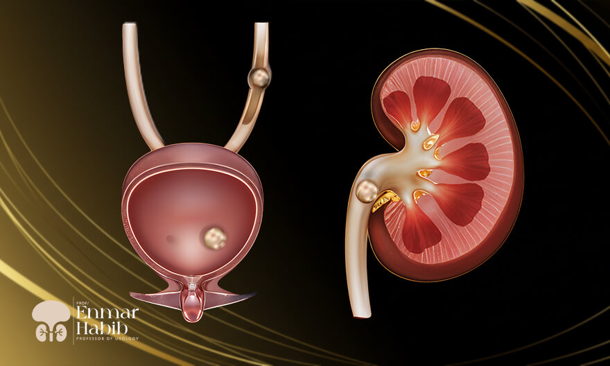
Stones are solid masses within the urinary tract and can cause pain, bleeding, infection, or obstruction of urine flow. Urinary tract stones can form in the kidneys and pass to the ureter or bladder. Depending on the location of the stone, it may be referred to as a kidney stone, ureteral stone, or bladder stone.
Dr. Enmar Mohamed Habib, Professor of Urology and Pediatric Urology at Cairo University and Fellow of McGill University, Canada, offers the latest techniques for urinary tract stone removal (kidneys, bladder, and ureters) without surgery. Some of the most well-known techniques include:
• Flexible ureteroscopy with suction technique.
• Minimally invasive nephroscopy with suction technique (percutaneous nephrolithotomy).
How do urinary tract stones form?
Urine contains many dissolved waste products. Typically, the kidneys remove these chemicals from the urine. In most people, having sufficient fluid helps flush them out. Additionally, other chemicals in the urine prevent stone formation.When there are too many waste products in a small amount of fluid, crystals begin to form. These crystals attract other elements and combine to create solid masses that grow larger unless they leave the body with urine.
Then, the stone may remain in the kidneys or travel through the urinary tract to the ureter. Sometimes, small stones depart the body in the urine without causing much pain. However, stones that do not move can cause urine to accumulate in the kidneys, ureter, bladder, or urethra, leading to pain.
Symptoms of urinary tract stones:
Some kidney stones may be as small as a grain of sand, while others may be the size of a small pebble or even much larger! Generally, the larger the stone, the more noticeable the symptoms. Kidney stones begin to cause pain when they lead to irritation or blockage. This pain can quickly progress into severe discomfort.Symptoms may include one or more of the following:
• Severe pain on either side of the lower back.
• Vague or persistent abdominal pain.
• Hematuria.
• Nausea or vomiting.
• Fever and chills.
• Foul-smelling or cloudy urine.
Causes of urinary tract stones:
Possible causes include drinking insufficient amounts of water, excessive or insufficient physical activity, obesity, weight-loss surgery, or consuming foods high in salt and sugar.If you have had one stone, you are at an increased risk of developing another. People who suffer from one stone have up to a 50% chance of developing another within 5 to 7 years.
Diagnosis of urinary tract stones:
Dr. Enmar Mohamed Habib, Professor of Urology and Pediatric Urology at Cairo University and Fellow of McGill University, Canada, begins the diagnosis of kidney stones with a medical history, physical examination, and imaging tests to determine the exact size and shape of the stones. A high-resolution computed tomography scan of the kidneys reveals the size and location of the stone.Kidney function is also assessed through blood and urine tests, along with an evaluation of the patient’s overall health and the size and location of the stone, to develop an appropriate treatment plan.
Treatment of urinary tract stones:
Dr. Enmar Mohamed Habib recommends drinking plenty of water to help the body pass small stones without additional procedures. However, if the stones are too large, obstruct the urine flow, or show signs of infection, they need fragmentation.1. Flexible ureteroscopy with suction technique:
Ureteroscopy is a minimally invasive procedure that involves passing a thin optical instrument called a ureteroscope through the urethra and bladder to reach the ureters and kidneys. The urethra is the tube from the bladder that drains urine. The ureter is the tube from the kidneys that carries urine to the bladder. The ureteroscope serves as a diagnostic and therapeutic minimally invasive method for the urinary tract, allowing precise access to the location of the stones. Ureteroscopy is essential for removing one or more stones from the kidneys that cannot pass through the ureter naturally. The treatment of upper urinary tract stones involves the use of a flexible ureteroscope assisted by holmium laser lithotripsy.What happens during the procedure?
1. X-rays are performed to confirm the presence of a stone.2. You are asked to refrain from eating or drinking for 6 hours before the surgery.
3. The procedure is performed under general anesthesia so that you remain asleep throughout the procedure.
4. The ureteroscope is inserted into the bladder and the affected ureter under X-ray guidance.
5. Using a probe or laser, Dr. Enmar Mohamed Habib then fragments the stones and removes them. Holmium laser lithotripsy is performed by inserting laser fibers that emit a light beam through the ureteroscope to reach the stone. The stone is then broken into smaller pieces or even dust, which is removed using a suction device.
6. A ureteral stent may be left in place.
7. A temporary catheter is placed and removed on the same day or the following morning.
8. After the procedure, you will be transferred to the recovery room and monitored until fully awake.
9. You can go home as soon as you can urinate normally. If you have a stent, a follow-up appointment will be scheduled for its removal.
What should I expect after the procedure?
• Drink plenty of fluids.• You may experience pain for at least 72 hours after the procedure.
• Take anti-inflammatory pain medications.
• You can return to work after a few days.
Advantages of the flexible ureteroscope:
• The flexible ureteroscope stands out from earlier devices by having an additional working channel (for irrigation or accessories such as lasers).
• Digital imaging ensures full clarity in the surgical field and allows for smaller equipment sizes.
• Access to the upper urinary tract without wall dilation or trauma.
2. Minimally Invasive Nephroscopy with Suction Technique:
What is a Nephroscope?
A nephroscope is used to examine the interior of the kidneys and treat certain upper urinary tract conditions without the need for traditional open surgery. It is an innovative, non-surgical treatment for large and complex kidney stones (typically stones larger than 2 cm) that cannot be passed through the urinary tract or may be difficult to treat using other kidney stone treatments, such as ureteroscopy. The nephroscope is inserted through the skin via a very small incision, allowing quicker recovery and reducing the need for post-operative drainage. The nephroscope contains internal channels that provide a light source, a viewing scope, and an irrigation system (a water system to wash the surgical site). Ultrasound or laser probes are used with the nephroscope to break down kidney stones. Once fragmented, the fragments are suctioned through one of the nephroscope's channels.How do I prepare for minimally invasive nephroscopy with the suction technique?
• A urine test will be performed, and you may need to take an antibiotic depending on the results.• If you are taking aspirin, stop it at least one week before the procedure.
• You should also discontinue any blood thinners and warfarin.
• You should not eat or drink for 8 hours before the procedure.
How is minimally invasive nephroscopy performed using the suction technique?
1. The best urologist, Dr. Enmar Mohamed Habib, reviews the latest tests (such as CT scans or urinary tract imaging) of the kidneys and urinary tract to prepare for the procedure.2. You will be given anesthesia, and a small catheter will be inserted through the urethra into the kidneys.
3. A dye is then injected into the catheter, and X-rays are taken to show details of the kidney's interior.
4. A needle is inserted through the skin of your back into the kidney at the exact location planned before the surgery. Through this renal access, a thin wire is passed into the kidney, and a balloon is used to dilate the area.
5. After dilation, the nephroscope and surgical instruments are inserted for direct access to the kidney. The stones are fragmented into small pieces so that they can be suctioned out using a suction device.
6. A ureteral stent is also placed in the kidney. The stent is a soft, hollow plastic tube that is placed along the entire ureter to keep it open, helping to drain urine and encouraging the kidney to heal. This stent is typically removed within a week after the procedure.
7. You will be transferred to the recovery room and monitored as you wake up from anesthesia to ensure there is no internal bleeding. Antibiotic treatment will continue.
8. You are likely to return home the day after the procedure.
9. The stones are sent for analysis to provide advice on preventing further stone formation.
What Are the Risks and Benefits of Nephroscopy?
Nephroscopy is a very safe procedure that reduces the need for traditional surgery, which involves longer recovery times and greater risks of injury.Benefits of Nephroscopy:
• High success rates, with most patients being free of stones 100% after the procedure.• Accurate localization of kidney stones and a better chance of completely removing them.
• Less pain after the procedure compared to open surgery.
• Fewer complications compared to open surgery due to the small incision and less invasive access to the kidneys.
• Faster return to daily activities and work compared to open surgery.
• Better rates of removing larger and more complex stones than less invasive options (ureteroscopy).
Ways to prevent kidney stones:
• Drinking enough fluids helps reduce the concentration of waste in the urine. Dark-colored urine is more concentrated, so your urine should be very light yellow to almost clear if you are well hydrated. Most people should drink more than 12 cups of water per day. If you exercise or the weather is hot, you should drink even more.• Most fluids you drink should be water, as water is better than soda, sports drinks, coffee, or tea.
• Eat more fruits and vegetables, which make the urine less acidic. When urine is less acidic, stones may be less likely to form. Animal protein produces urine that contains more acid, which can increase the risk of developing kidney stones.
• Reduce excess salt in your diet. Avoid foods high in salt, such as salty potato chips, French fries, canned meats, canned soups, packaged meals, and even sports drinks.
• If you are overweight, work toward reaching a healthy weight. However, high-protein weight-loss diets may increase the risk of kidney stones. You need enough protein, but as part of a balanced diet, to lower the risk of stone formation.
Modern techniques for removing urinary tract stones, such as flexible ureteroscopy and minimally invasive nephroscopy with suction technique, are effective methods that provide non-surgical solutions to kidney and ureter stone problems. By utilizing these advanced procedures under the supervision of Dr. Enmar Mohamed Habib, Professor of Urology and Pediatric Urology at Cairo University and Fellow of McGill University, Canada, patients can receive precise and safe treatment, contributing to faster recovery and reduced complications. Therefore, if you are experiencing urinary tract issues, do not hesitate to consult Dr. Enmar Habib for an evaluation of your condition and to receive optimal solutions.
خدماتنا
بفضل التقدم في التقنيات الجراحية، أصبح يمكن علاج ضيق مجرى البول بالتدخلات المحدودة والتعافي السريع باستخدام الهولميوم ليزر.
يعد السلس البولي لدى الأطفال مسألة صعبة ومحرجة لكل من الأطفال والآباء، ويتطلب العلاج تحديد الأسباب بدقة لوضع خطة علاجية مناسبة.
يكمن العلاج الفعال لمشاكل الجهاز التناسلي للأطفال في دقة التشخيص، لذلك يجب معرفة الفرق بين الخصية المُعلقة والخصية المُرتجة.